Invented by Hyun Hwa Oh, Young Hun Sung, Jae Hyun Kwon, Kang Eui Lee, Samsung Electronics Co Ltd
Medical imaging has become an integral part of modern healthcare. It is a non-invasive diagnostic tool that helps doctors to visualize the internal structures of the human body. Medical imaging apparatus, such as X-ray machines, CT scanners, MRI machines, and ultrasound machines, are used to capture images of different parts of the body. These images are then displayed on a monitor for doctors to analyze and diagnose medical conditions.
The market for medical imaging apparatus to display X-ray images from different types of medical imaging modalities is growing rapidly. The demand for medical imaging equipment is driven by the increasing prevalence of chronic diseases, such as cancer, cardiovascular diseases, and neurological disorders. The aging population is also a major factor contributing to the growth of the market.
X-ray imaging is one of the most commonly used medical imaging modalities. It is a non-invasive procedure that uses electromagnetic radiation to capture images of the internal structures of the body. X-ray machines are used to diagnose a wide range of medical conditions, such as bone fractures, lung infections, and dental problems.
The market for X-ray imaging apparatus is expected to grow at a CAGR of 6.2% from 2020 to 2027. The increasing demand for digital X-ray systems, the growing number of diagnostic imaging centers, and the rising prevalence of chronic diseases are the major factors driving the growth of the market.
CT scanners are another type of medical imaging apparatus that is used to capture images of the internal structures of the body. CT scans are used to diagnose a wide range of medical conditions, such as cancer, cardiovascular diseases, and neurological disorders. The market for CT scanners is expected to grow at a CAGR of 5.7% from 2020 to 2027.
MRI machines are also widely used in medical imaging. They use a strong magnetic field and radio waves to capture images of the internal structures of the body. MRI machines are used to diagnose a wide range of medical conditions, such as brain tumors, spinal cord injuries, and joint problems. The market for MRI machines is expected to grow at a CAGR of 5.3% from 2020 to 2027.
Ultrasound machines are another type of medical imaging apparatus that is used to capture images of the internal structures of the body. Ultrasound machines use high-frequency sound waves to create images of the internal structures of the body. They are used to diagnose a wide range of medical conditions, such as pregnancy, heart problems, and liver diseases. The market for ultrasound machines is expected to grow at a CAGR of 6.5% from 2020 to 2027.
In conclusion, the market for medical imaging apparatus to display X-ray images from different types of medical imaging modalities is growing rapidly. The increasing prevalence of chronic diseases, the aging population, and the growing number of diagnostic imaging centers are the major factors driving the growth of the market. X-ray machines, CT scanners, MRI machines, and ultrasound machines are the most commonly used medical imaging apparatus. The market for these machines is expected to grow at a CAGR of 5.3% to 6.5% from 2020 to 2027.
The Samsung Electronics Co Ltd invention works as follows
Disclosed are a display device that allows simultaneous comparison of multiple images, each illustrating different features on a divided screen. The apparatus also provides an image display method that is possible by using the apparatus. The display apparatus comprises a memory that can store a variety of images of an object, and an input device that can receive a command to simultaneously display the different types. The command is received by the display device. It divides the screen into two regions. One region displays a portion of an object’s image, while the second region displays a second image.

Background for Medical imaging apparatus to display x-ray images from different types
1. Field
Exemplary embodiments refer to a display device that is usable for showing images.
2. “2.
Comparing images is often necessary when images are viewed using display devices. Comparing images is sometimes necessary in medicine where multiple medical imaging apparatuses are used to identify lesions. The images that need to be compared are generally displayed on multiple display devices in order to check the results of multiple images taken with different types of medical imaging apparatuses.
It may be more difficult to quickly compare images from the same region of interest when multiple display devices are being used than if one apparatus is used. It is possible for a user to lose focus or become distracted by the multiple display apparatuses that must be used to check the region of interest.
Therefore, one or more of the exemplary embodiments provides a display device that allows simultaneous comparisons of multiple images on a split screen. The image display method is also possible by using this apparatus.
Additional features of the exemplary inventions will be described in part in this description. In part, the description will be obvious or can be learned through the practice of the example embodiments.
According to one aspect of one of many exemplary embodiments, a device for displaying images includes a memory that can store a plurality different types of images of an object, an inputting device which can receive a command relating to simultaneously displaying these different types of images and a display device which is configured to display images. The display device further comprises a screen on which an image of an object is displayable. A first region displays a portion of the object and a second area displays a second image.
According to another aspect, one or more exemplary embodiments of a display apparatus comprises a memory configured for storing a plurality different types of images of an object and an input device configured receive an input of one between a division command or a shift command. A screen on which an image is displayed of the object can be divided into a first area in which a first picture showing one portion of it is displayed and a region in which a second photo showing the remaining portion is displayed. The display device can also change the proportional amounts of each image and the respective portion of the objects shown in the first and the two images based on the respective percentage of the respective portion of the respective amount of the respective portion of the second images
According to another aspect of one of more exemplary embodiments, an imaging display method that is executable using a display apparatus involves receiving an input of screen division commands which relate to dividing a screen. A first region in which a portion of an object’s image is displayed is divided into a region where a portion of the object’s image is displayed, and a second area within which a portion of the object’s second image is displayed. Based on the screen division command.

According to a further aspect, one or more exemplary embodiments of an image display system for displaying a plurality images showing a particular region of an object involves seamlessly displaying one of each of these images on a device so that the displayed portions are combined to create a combined image of the object’s specific region.
Reference will now take place in detail to the exemplary embodiments. Examples of these are shown in the accompanying illustrations. Like reference numerals refers to like elements throughout.
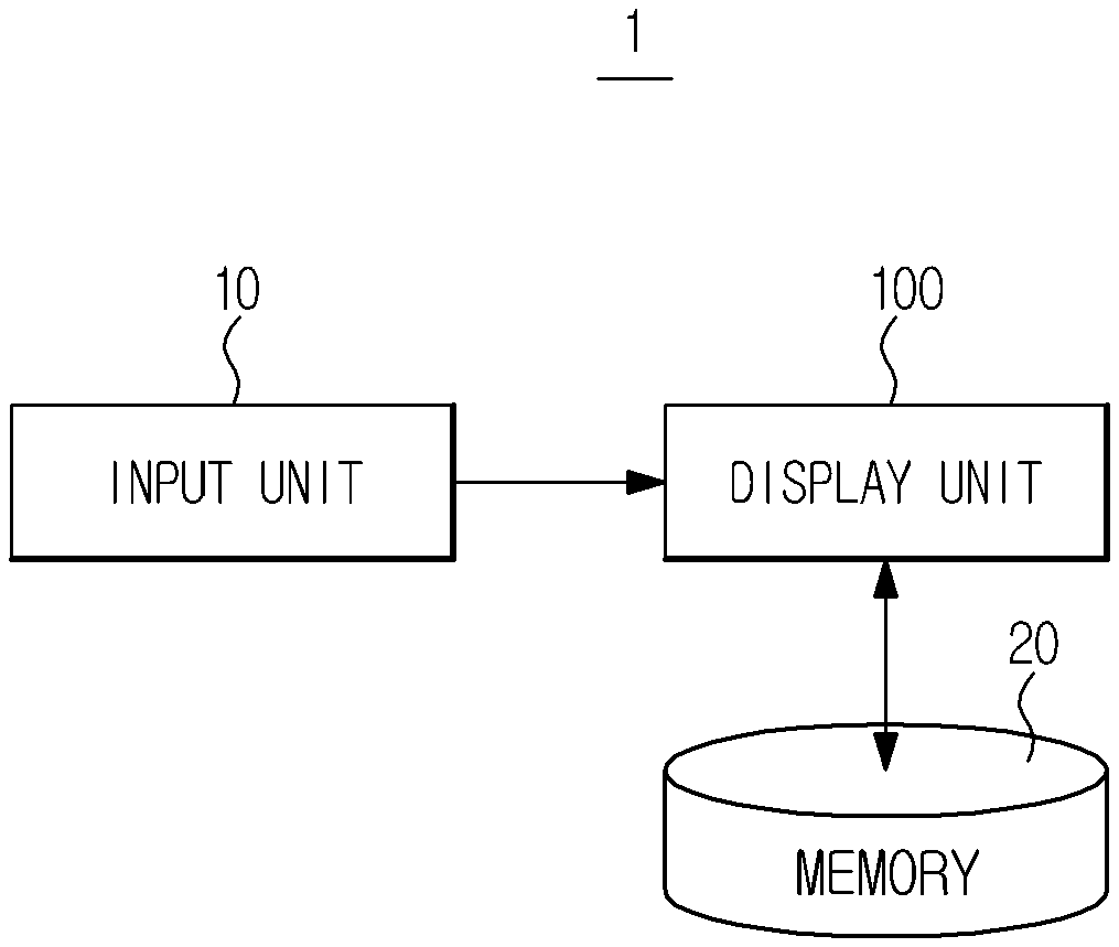
FIG. “FIG. Display apparatus 1 contains a display unit, also known as a “display device” Display unit and/or display? 100 used to display an image. A memory 20 is used to store images of objects. An input unit (also known as an ‘input device?) is also used to display the object. 10 for receiving an input of a command relating to manipulation of the display apparatus. An exemplary embodiment of the display apparatus 1 includes any of the following devices that can display images: a tablet, a smartphone, a laptop, or a tablet.
The display unit 100 is a component of the display apparatus 1. It includes a screen that displays an image. However, it can be implemented using any of many different display techniques. The touchscreen can be used to allow a user to touch the display 100 directly or via the input device 10. You can input any command to the display apparatus 1 using a finger, touch pen, or stylus. One or more of the input units 10 can include a keyboard, mouse, joystick, track ball, joystick, voice recognition device and motion recognition device. The input unit 10 can be either integrated into the display apparatus 1 (or installed within the display apparatus 1) The input unit can also be supplied separately from the display device. If the input unit is not connected to the display device, it may transmit a received command via wireless communication to the display unit 10. It may also be connected to the apparatus using any of a variety of connectors. Sometimes, the user may need to inspect images of the same object or a particular region of an object using the display apparatus 1. This is especially important in medicine. A variety of modalities can be used in medicine to diagnose disease. These include an x-ray machine, an ultrasound apparatus and a computed radiography apparatus. To photograph an object, each modality may use one or more different imaging techniques. The xray apparatus can obtain a variety of imaging images, including a general image that shows all bones and soft tissue, a soft-tissue xray image showing only organs and a color image showing a stark contrast of colors.
The medical staff compares medical pictures of an object that were captured using different techniques and modalities to examine a suspected lesion. Multiple display devices are used to compare different types of medical images. Multiple display devices are often used to compare different types of medical images. However, a single device can be more efficient and intuitive. The disclosed embodiments include a user interface that allows simultaneous comparison and checking of multiple medical images captured using different techniques. In this context, the exemplary embodiments are described with respect to medical diagnosis x-ray images. The technical spirit of the exemplary inventions is not restricted to medical images. It is applicable to all fields that require the analysis and comparison of multiple images.
FIG. “FIG. As examples of xray images, 3 includes a general (general), soft tissue (soft) and bone (bone) xray images.
Referring to FIG. 2. A captured x-ray image showing the chest region of an object can be seen on the display unit 100. Markers 110 and 111, which can be used to divide the screen, are displayed respectively on the left and right side of the display 100. An example of a medical image is the x-ray. The display unit 100 can display medical images. These images are not restricted to x-ray images. They also include images captured using other types of modalities such as an ultrasound apparatus or a computed imaging apparatus, magnetic resonance imaging apparatus, positron emission apparatus, and single photon emission computedtomography apparatus.
Drawing (a), FIG. 2. This is a view of the display area 100 before a screen division was implemented. The entire display area of the display unit 100 is made up of a first region 120. This displays a first image 121, which corresponds to an x-ray image. The first image 121 is displayed in the 120th region prior to the implementation of a screen division. It can be used as a reference image and compared to other images that have been taken using different techniques. The general x-ray image in FIG. 3. may be used as the first image 121. These examples are only an example. There are many types of images that can be captured by different techniques. For example, bone xray images, soft tissue images, and/or color xray images of the same object as shown in FIG. A user may choose to display the 3 images.
As shown in FIG. 2 (b), the user clicks on the marker 110, which is located on the left side display unit 100. 2 and the user is prompted to drag the marker 110 to its right side, as shown in FIG. The screen is divided into two regions: the first 120 and the second 130. Clicking the marker 110 creates a vertical boundary line, vb1, which runs through the marker 110. The marker 110 can be dragged so that the vertical boundary line moves with it. The vertical boundary line (vb1) serves as a boundary between two regions: the first 120 and the second 130.
As shown on FIG. 2. The direction that the marker 110 can be moved may be indicated with a symbol such as an arrow. Clicking and dragging the marker 110 can also be done via the previously mentioned input unit 10, which could include a mouse, keyboard, joystick, joystick, jog wheels, voice recognition devices, and motion recognition devices.

As shown on drawing (c), FIG. 2. When the vertical boundary line (vb1) is moved to the left side by clicking on the marker 110 and dragging it to the right of the display unit 100 the screen share for the first 120 regions decreases and the screen share for the second 130 regions increases. The screen share of first region 120 drops, and the proportion of chest area of object in the first image 121 shown in the first regional 120 also decreases. FIG. 2. The entire object is shown in the first image 121. Drawing (c) in FIG. 2. The first image shows only the right-hand side of the object’s chest. The second image 131 displays the left-hand portion of the chest in the second region 130. The entire chest area of the object is represented by the combination of the first 120 region, in which the first image is displayed, and second 130 region, in which the second image is displayed.

Click here to view the patent on Google Patents.