Invented by MAVROPOULOS; Anastasia, LUENGO HENDRIKS; Cristian L., ZHANG; Senzeyu, CARELLI; Ryan C., JACOBS; Kevin B., DEEPCELL, INC.


This article will help you understand a new patent application that changes how we look at dying cells. Using machine learning and computer vision, this invention makes it possible to study cells without using labels or stains. Let’s explore what this means, why it matters, and how it works in detail.
Background and Market Context
Cell death is a key process in biology. Every living thing has cells that live, get sick, and die. When cells die in the body, it can mean normal growth, healing, or disease. For years, scientists have wanted to know how and why cells die. They study this to learn about cancer, infections, and how drugs work.
Traditionally, researchers use stains or dyes to see which cells are dying. These dyes stick to certain parts of a cell or react with certain proteins. For example, Annexin V sticks to a cell’s surface during early cell death called apoptosis. SYTOX™ dyes can show up when the cell’s membrane breaks during late stages of cell death. These tools are helpful, but they have problems. Stains can change how a cell looks or works. Some dyes kill the cells or make it hard to use the cells for other tests. Also, these methods can be slow and need a lot of work.
To speed things up, labs use machines like flow cytometers. These machines can count and sort cells quickly, but they still need stains or other labels. This means scientists can miss important types of cells if they don’t have the right label. Sometimes, interesting cells are not seen at all because they don’t have the expected marker.
The need for faster, less damaging, and more accurate cell analysis is growing. Drug companies, hospitals, and research labs all want better ways to study cell death. They want tools that are quick, cheap, and that let them keep working with the cells after they are analyzed. That’s where this new invention comes in.

This patent describes a method using machine learning and computer vision. It can study dying cells without staining them, making the process easier, safer for the cells, and more reliable. This is important for fields like drug discovery, cancer research, and even personalized medicine, where understanding how cells die can guide treatment.
Scientific Rationale and Prior Art
To understand this invention, it helps to know how scientists have studied cell death in the past. For many years, the main way to tell if a cell is dying was to use chemical stains or dyes. These stains react with certain molecules inside the cell. For example:
– Annexin V binds to phosphatidylserine, a molecule that moves to the outside of the cell during early cell death.
– SYTOX™ dye enters cells that have lost their membrane, showing late-stage death.
These stains are seen under a microscope or with machines that use lasers to detect the color. Scientists look at the colors and decide if a cell is healthy, dying, or dead. Sometimes, other chemical reactions or enzymes are used as markers.
But there are big limits to these ways. Staining can kill the cell or change what the cell looks like, so you can’t use the cell for other tests. Also, stains only show a few features, so you might miss important changes. Flow cytometry machines can sort cells using these stains, but again, only if you have the right stain for the right feature.
Another way to study cells is to look at their shape and size. This is called morphology. Scientists use microscopes and try to describe what they see, but this is slow and can be hard to compare between people. Computers can help by measuring things like size, roundness, or how dark or light the cell is in a picture. This is called morphometric analysis.


In recent years, scientists started using machine learning, especially deep learning, to find patterns in pictures of cells. These computer programs can “see” very small details that humans might miss. They can be trained using thousands or millions of pictures, learning to spot patterns linked to different kinds of cell death.
Before this invention, some groups tried using computer vision or machine learning to study cells, but usually they still used stains, or they only looked at a few features. No one had put together a system that could look at unstained, dying cells, measure many features at once using both machine learning and standard image analysis, and link those features to the type or stage of cell death. This is what makes the new patent different.
Invention Description and Key Innovations
This invention is a method and system for studying dying cells using label-free imaging, machine learning, and computer vision. Here’s how it works in simple terms:
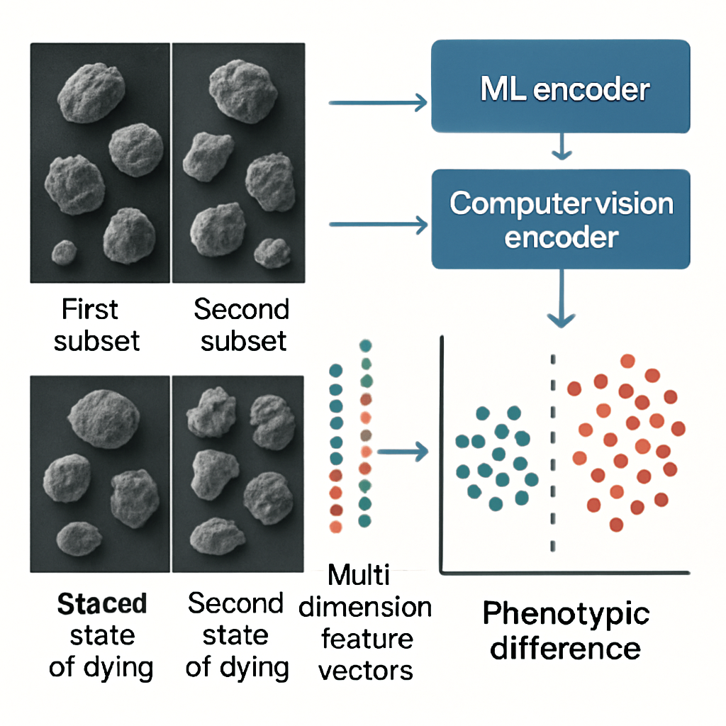
First, cells are put into a special channel, like a very small tube, called a microfluidic chip. The cells flow through this channel, one at a time, in a thin stream. As each cell passes by, a high-speed camera takes a picture of it. These pictures are called brightfield images, which means the cells are seen in black and white, using normal light.
The special thing is that the cells do not need to be stained or labeled in any way. The picture is enough.
Next, each picture is sent to two types of computer programs:

1. A machine learning encoder (often a deep neural network) looks at the image and pulls out features. These might be things like patterns, textures, or other details that are hard for humans to describe. These features are called “ML-based features.” They are usually just numbers that show how much of a certain pattern the computer found.
2. A computer vision encoder uses rules made by people to measure things that humans can understand, like the cell’s size, shape, roundness, and how bright or dark it is. These are called “morphometric features.” For example, the program might measure the area of the cell, how long and wide it is, or how smooth its edge is.
Both sets of features are put together into a long list of numbers, called a multi-dimensional feature vector. Each number in the list stands for a different feature. This vector is like a fingerprint for the cell—it sums up everything about how the cell looks.
The system then uses these vectors to group cells. It can tell which cells are in one stage of dying and which are in another. For example, it might notice that some cells are shrinking, some are breaking apart, and some are swelling. By comparing the vectors, the system can sort cells into groups: healthy, early dying, late dying, or dead.
This sorting can be done in real time. As the cells flow through the channel, the computer decides, based on the fingerprint, which cells should go into which collection tube. This is useful for researchers who want to save certain cells for more tests.
The key innovations here include:
– The ability to study and sort dying cells without any stains or labels.
– The use of both machine learning and rule-based computer vision to get a very complete picture of each cell.
– Real-time analysis and sorting, so the process is fast and the cells can be used for more experiments.
– The system can work with many types of cell death, like apoptosis (planned cell death), necrosis (unplanned, messy death), or other special types.
The machine learning part is trained using millions of cell pictures, so it gets very good at spotting small differences. The computer vision part adds features that scientists can understand and check.
The system can also reduce the complexity of the data. If each cell has 100 features, it might be hard to look at all at once. The computer can use math (like UMAP or PCA) to show the data in just two or three dimensions. This makes it easy to plot and see how cells group together.
The method is simple for the user. You put in your cell sample, the machine takes pictures, the computer analyzes them, and you can sort or study the cells based on their state of death. The cells are not harmed by stains or dyes, so you can do more tests on them later.
This invention makes cell death analysis easier, faster, and more flexible. It opens up new ways to study how drugs affect cells, how diseases like cancer progress, and how the body responds to different treatments. Because it doesn’t need labels, it can find rare or unusual cell types that would be missed with normal methods.
Conclusion
This new patent changes how scientists study dying cells. Instead of relying on stains or human judgment, it uses machine learning and computer vision to analyze cells in their natural state. This means more accurate, faster, and less damaging cell analysis. The system can sort and save cells for more tests, and it gives a much deeper look at the process of cell death. As more labs use this technology, we will learn much more about health, disease, and how to create better treatments.
Click here https://ppubs.uspto.gov/pubwebapp/ and search 20250218201.