Invented by Behzad Sharif, Debiao Li, Daniel S. Berman, C. Noel Bairey Merz, Cedars Sinai Medical Center
Myocardial perfusion MRI is a non-invasive imaging technique that allows doctors to assess the blood flow to the heart muscle. This technique is particularly useful in diagnosing heart disease, as it can detect areas of the heart that are not receiving enough blood. However, traditional myocardial perfusion MRI requires electrocardiogram (ECG) gating, which can be time-consuming and uncomfortable for patients.
To address this issue, several companies have developed systems and methods for myocardial perfusion MRI without the need for ECG gating. These systems use advanced imaging techniques to capture images of the heart at different stages of the cardiac cycle, allowing doctors to assess blood flow without the need for ECG gating.
One such system is the Siemens Healthineers MAGNETOM Vida MRI system, which uses a technique called Free-Breathing Myocardial Perfusion (FBMP) to capture images of the heart without the need for ECG gating. This system uses advanced motion correction algorithms to compensate for the motion of the heart during the cardiac cycle, allowing for high-quality images of the heart at rest and during stress.
Another system is the Philips Ingenia Elition MRI system, which uses a technique called Compressed SENSE to capture images of the heart without the need for ECG gating. This system uses advanced image reconstruction algorithms to create high-quality images of the heart in a fraction of the time required for traditional ECG-gated myocardial perfusion MRI.
In addition to systems for myocardial perfusion MRI without ECG gating, there are also additional systems and methods for image reconstruction and analysis. These systems use advanced algorithms to analyze the images captured during myocardial perfusion MRI, allowing doctors to assess blood flow to the heart and identify areas of the heart that may be at risk for heart disease.
One such system is the HeartFlow Analysis System, which uses advanced computational fluid dynamics algorithms to create a 3D model of the coronary arteries and assess blood flow to the heart. This system can help doctors identify areas of the heart that may be at risk for heart disease and develop personalized treatment plans for patients.
Another system is the Circle Cardiovascular Imaging cvi42 software, which uses advanced image analysis algorithms to analyze images captured during myocardial perfusion MRI. This software can help doctors assess blood flow to the heart and identify areas of the heart that may be at risk for heart disease.
Overall, the market for systems and methods for myocardial perfusion MRI without the need for ECG gating, and additional systems and methods for image reconstruction and analysis, is growing rapidly. These systems and methods are helping doctors diagnose and treat heart disease more effectively, and are improving the quality of life for patients with heart disease. As technology continues to advance, we can expect to see even more innovative systems and methods for myocardial perfusion MRI in the future.

The Cedars Sinai Medical Center invention works as follows
In certain embodiments, the present invention discloses systems and methods of cardiac MRI that permit continuous un-interrupted acquisition with no ECG/cardiac gating/synchronization that achieves imaging perfusion defects. An accelerated image reconstruction method is also described in the invention. This technique can be tailored to the data acquisition scheme, and minimizes or eliminates dark-rim artifacts. Concurrent imaging of perfusion and myocardial motion (cardiac function) is possible through the invention. This can reduce exam time and provide additional diagnostic information for CAD patients.

Background for Systems and Methods for Myocardial Perfusion MRI Without the Need for ECG Gating, and Additional Systems and Methods for Improved Cardiac Imaging
The following description contains information that may help you understand the invention. This description does not imply that any information contained herein is of prior art or relevant for the claimed invention.
A decrease of stress myocardial blood flow is an early indicator of functional effects related to abnormalities in the heart. Myocardial perfusion imaging is (MPI) the most common method of assessing it. CMR is still not widely used, especially in the United States. However, multiple multicenter trials have shown higher detection rates for CAD with CMR than with MPI. Vasodilator stress CMR CMR first-pass imaging has been improved with recent hardware and software upgrades. This allows for comprehensive cardiac assessment using a radiation-free method.
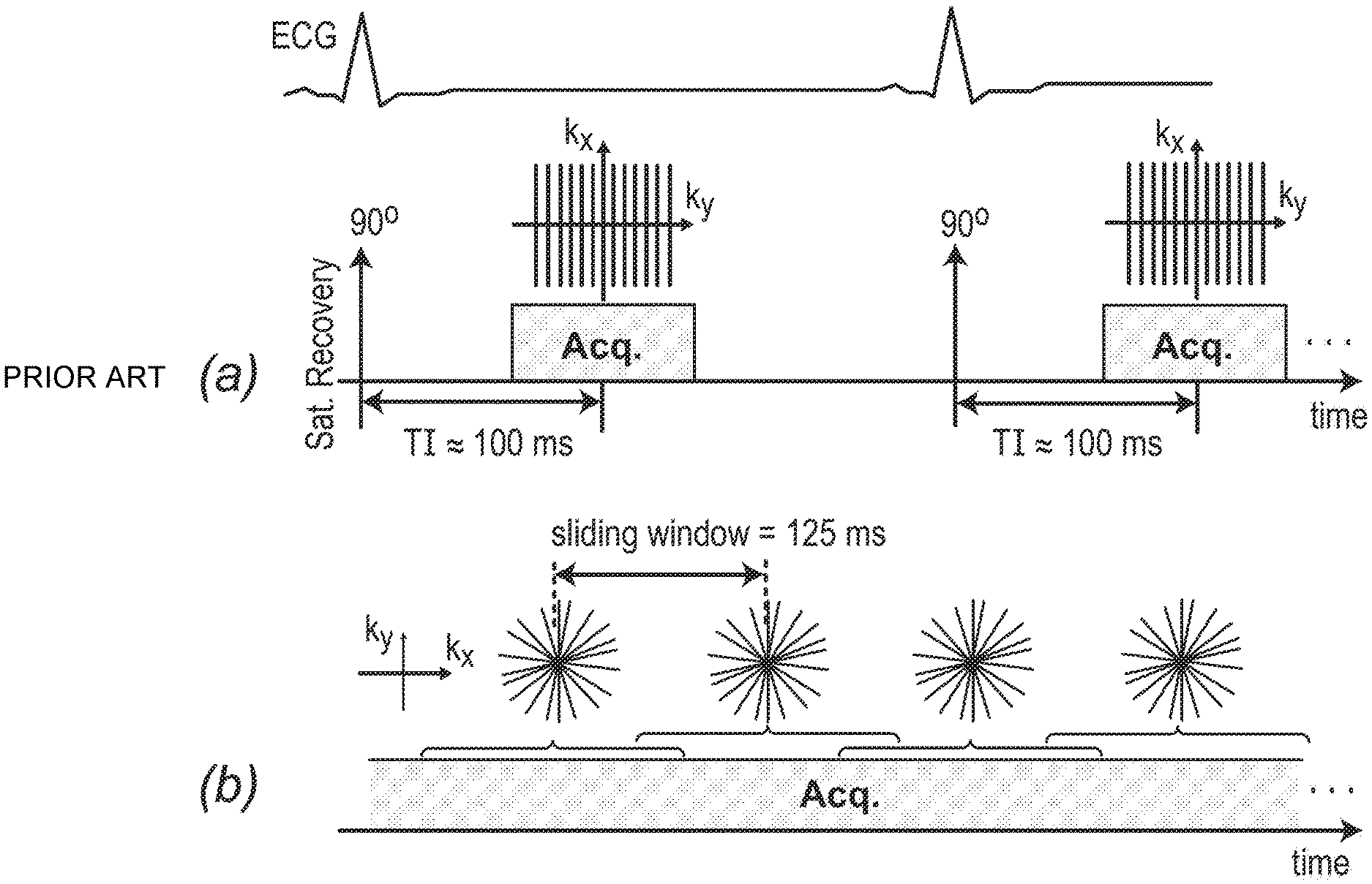
Despite the many technical advancements made over the past decade, perfusion CMR has been limited in widespread use. CMR is a complicated method that is more dependent on the technical expertise of the technician than nuclear myocardial orfusion imaging. It is also considered a difficult modality to diagnose CAD. Complexity has also been caused by the requirement for ECG gating that is nearly perfect.
Another problem is stress MRI studies often include an artifact that makes image interpretation challenging even for experts. The subendocardial dark rim artifact refers to this image artifact. Dark-rim artifacts in the perfusion image series are more prominent when the contrast bolus enters the left ventricular cavity. This is especially true during the stress portion. This artifact mimics true perfusion defects and lasts only a few beats. Dark-rim artifacts are still a problem for perfusion MRI accuracy and widespread adoption. They impede the diagnosis of hypoperfusion (subendocardium), which is the layer of myocardium that is most commonly seen to be abnormal in ischemic heart disease.
CMR stress perfusion imaging offers the promise of a complete cardiac exam without radiation. The high resolution of CMR allows for the identification of subendocardial and subepicardial hypoperfusions, which cannot be solved by nuclear methods. This perfusion CMR quality could solve the problem of?balanced decrease of flow? This is a known mechanism through which nuclear MPI can miss high-risk forms of the disease.
As a result of the above considerations, there is a clear need in the art of a CMR perfusion technique which eliminates ECG gating and produces images without the dark-rim artifact.
In various embodiments, this invention teaches a method to perform first-pass magnetic resonance imaging (MRI) in myocardial perfusion. In some embodiments, the method includes: (1) using an MRI machine to apply a pulse sequence with radial k-space sampling to a volume of interest (VOI) including a region of a subject’s heart; (2) introducing a contrast agent into the subject’s vascular system prior to or during imaging; and (3) using a model-based/iterative image reconstruction technique to generate one or more images of one or more slices or VOIs within the heart region. In some cases, MRI is not required to acquire an electrocardiogram (ECG). An apodization scheme may be used in image reconstruction to reduce or eliminate dark-rim artifacts. One or more images may depict the heart, or a portion thereof, within 30-60 heartbeats following injection of contrast agent. Multiple frames are created per heartbeat in certain embodiments at an average of 8 frames per second. This allows for the depiction of heart movement. An apodization scheme can be used in some embodiments to reduce or eliminate dark-rim artifacts. The MRI machine can be a 3.0T scanner in certain embodiments. Some embodiments may have an arrhythmia.
In different embodiments, the invention teaches a technique for performing cardiac magnetic resonance imaging. The method can be used to (1) apply an ungated gradient recalled Echo (GRE), pulse sequence with continuous radial capture to a volume or interest (VOI), and (2) use an iterative parallel imaging technique or sensitivity encoding picture reconstruction technique to generate images of one or several anatomical structures within that VOI. The method may also include injecting a contrast agent into a subject’s bloodstream prior to or during imaging. An apodization scheme can be used in image reconstruction to reduce or eliminate dark-rim artifacts. The MRI machine can be a 3.0T scanner in some embodiments. Certain embodiments may be used when the subject suffers from arrhythmia. Multiple image frames are generated for each heartbeat in some embodiments at a rate at least 8 frames per seconds to show the heart motion. The method may also include diagnosing the subject based on one or more images.
The invention teaches a method of performing cardiac magnetic resonance imaging. The method may include (1) using an MRI machine with continuous radial sampling to a volume in interest (VOI), wherein the VOI is a region of the subject?s heart; (2) injecting a contrast agent into the subject?s vascular system before or during imaging; (3) and using an iterative image reconstruct scheme to generate images of one or several anatomical structures within that VOI. An apodization scheme can be used in image reconstruction to reduce or eliminate dark-rim artifacts. Multiple image frames are generated for each heartbeat in some embodiments at a rate at least 8 frames per seconds to show the heart movement. In some embodiments, the method also includes diagnosing the subject based on one or more images. The MRI machine can be a 3.0T scanner in certain embodiments. Certain embodiments may cause arrhythmia in the subject.
The invention describes a magnetic resonance imaging method in various embodiments. The system may include (1) a magnet that can generate a magnetic field, (2) a transmitter capable of transmitting to a region within that magnetic field, (3) a receiver capable of receiving a magnetic resonance signal in the region, and (4) a processor which is able to control the transmitter or receiver. In some embodiments, the processor is able to direct the transmitter to send a sequence to the receiver and transmitter, including (a), applying any of these pulse sequences to a volume (VOI), in which the subject’s heart region of the magnetic resonance data; (VOI); and (c) creating an image using the processor to create a image based on the data.

In various embodiments, this invention teaches a machine-readable medium that contains machine executable instructions to cause one or more processors to a magnetic resonance imaging machine (MRI) to execute a method. This includes: (1) applying any one of the pulse sequences herein to a subject’s volume of interest, wherein the VOI is a region in the subject’s chest; (2) obtaining magnetic resonance data from the subject’s volume of interest; (3) generating one (or more) images based upon the magnetic resonance data, using any imaging generating methods.

Click here to view the patent on Google Patents.